BASC IMAGING
BASC Imaging is an independent, low cost, non-hospital imaging facility featuring true Open MRI technology, Low dose CT Scans and Ultrasound services. We are the Radiology Department of the Bedford Ambulatory Surgical Center (BASC) that serves our community and providers with the same level of excellence known for over 30 years.

ONLINE BILL PAY
Online bill pay is here to make for quick and easy bill paying for all of our facilities. Click on the button below to get started.
Frequently Asked Questions
We want to make your experience at the BASC Imaging as easy as possible, so we’ve compiled some of our most frequently asked questions and answers below. If you still have questions, give us a call and our team will be happy to help you.
IMAGING INFORMATION
Once the report is completed, a copy will be faxed within 24 – 48 hours to the ordering provider’s office. Patients are encouraged to call their ordering provider to obtain a copy of their report prior to calling BASC Imaging. Patients can request a copy of their report by calling 603-296-0723 or coming to BASC Imaging and picking up a copy. You can email info@bascimaging.com to request a copy as well. Please be aware patients requesting their report will be available 7 – 10 days after exam.
Anything is acceptable to wear for CT/Ultrasound. MRI patients should remove clothing that contains metal such as pants with zipper/buttons, underwire bras, etc. We will provide shirts and shorts if needed and a locker for you to secure your valuables.
All imaging orders should be faxed to 603-232-0056 or emailed to info@bascimaging.com
Once we receive an order from the ordering provider with all necessary information including insurance authorization, we will call the patient to schedule an appointment.
Patient images can be sent electronically through a secure portal to most provider practices. Images can also be sent to the provider by mail at the request of the patient. Patients can request a CD of their images at the time of their appointment as well.
BASC Imaging utilizes two radiology groups. Southern New Hampshire Radiology Consultants (SNHRC), is a physician-owned professional corporation providing Diagnostic Imaging Services to patients in the greater Manchester, NH area.
SNHRC is comprised of a group of highly trained, board-certified radiologists, with fellowship trained sub-specialists in the areas of Neuroradiology, Body Imaging, Musculoskeletal Radiology, Pediatric Radiology, Interventional Radiology and Breast Radiology. www.snhrc.com
We also utilize New England Baptist Hospital Radiology Group. All exams are interpreted by fellowship-trained, sub-specialty radiologists who also have expertise in musculoskeletal ultrasound for both diagnostic and therapeutic applications. www.nebh.org
CT Scans will be scheduled for 15-30 minutes. MRI’s will be scheduled 30-60 minutes. Ultrasound will be scheduled for 30-45 minutes. Scan length time depends on the type of exam being performed.
YES! By law you have a CHOICE where to go for your imaging exam. Let your provider know you REQUEST to go to BASC Imaging. Regardless of where your imaging has been done in the past such as a hospital, BASC Imaging is happy to accommodate your imaging request.
BASC Imaging is happy to assist you in identifying the estimated cost for your specific scan and insurance. By choosing BASC Imaging over Hospital based facilities, patients are able to save significant amounts of money, which can be helpful if you have a large deductible or no health insurance at all.
Call us at 603-296-0723 or Email at info@bascimaging.com
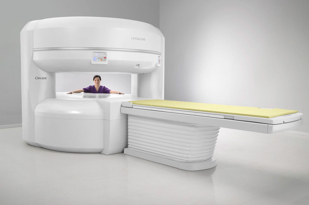
OPEN MRI
Our MR scanner can accommodate patients weighing up to 660lbs. The opening is approximately five feet across, so the person will need to be able to fit inside that area.
Inside the scanner are coils of metallic wire. When electricity passes through the coils, a magnetic field is created and the coils vibrate. The sound the MRI makes is the metal coils vibrating and banging together inside the machine, creating a very strong magnetic field. We provide ear plugs for protection and have a noise cancellation sound system so the patient may enjoy music through headphones while being scanned.
Pacemakers and defibrillators cannot go into an Open MRI unit. Stents, aneurysm clips, non-removable electronic devices, loop recorders and metallic foreign bodies need to be reviewed by the Radiologist prior to the exam.
If you have any form of implant or have sustained an injury where it was confirmed or suspected that you may have shrapnel in your body, please inform BASC Imaging when scheduling your appointment.
Make and model # of implant are required. Some implants are able to be placed in an Open MRI while others are conditional to a Closed MRI only.
In order to obtain optimum image clarity from your MRI scan, you will need to hold still. The MRI technologist will inform you when you can move between scans. We aim for the best quality scan for you and your provider.
In certain circumstances, we are fine with family members and loved ones to accompany the patient in the room as long as they pass the screening qualifications. The MRI Technologists have a lot of experience comforting and reassuring patients before and during the scan.
MRA, or Magnetic Resonance Angiogram, is a type of MRI scan that takes images of blood vessels in the body. MRA scans are often used to evaluate for an aneurysm, blocked arteries and evaluate levels of stenosis. MRA’s can provide information that cannot be obtained by x-ray, ultrasound, or CAT scans.
Open MRIs allow a spacious scan to take place, often alleviating any claustrophobic tendencies patients might experience with a tightly enclosed MRI. Our open MRI machine has open sides, allowing for a clear, unobstructed view of your surroundings. Open MRI machines often are more accommodating for most size individuals.
Closed MRI units are traditionally tunnel-style. This tube environment often causes claustrophobic prone patients distress during their long scan periods. In addition, most closed MRI machines are impossible for larger patients to use.
MRI, or Magnetic Resonance Imaging, uses a large magnetic field and radio frequency pulses to create images in any plane. MRI’s produce detailed images of organs, soft tissue, tendons, ligaments, the spinal cord, bone, and other internal body structures. The detail between normal and abnormal tissue is clearer on an MRI image than a CT scan. MRI’s can also generate 3D images of the vessels in your body. There is no radiation with a MRI scan.
A CT scan, or Computed Axial Tomography, uses x-rays for imaging. CT scans are obtained much quicker than MRI, but provide less detail. MRI patients who are unable to have an MRI due to safety reasons (metal implants, etc) their provider may order a CT scan instead. CT scans are quick (about 10 minutes) while an MRI scan is 30 – 45 minutes.
MRI contrast is a rare earth mineral (Gadolinium). Our body already has this mineral, but by injecting more into the blood stream, we are able to differentiate between normal and abnormal tissue much more accurately. The contrast does filter through your body, so it is important that your kidney function is of an acceptable level before administering any contrast to your system.
The contrast we use is extremely safe, however, there are some risks factors which will be discussed at time of scheduling.
In order to get the best images possible, the part of the body being studied has to be in the middle of the scanner. Thus, if you are having a brain MRI, your head will have to be in the middle of the scanner. If you are having an ankle MRI, your ankle will be in the scanner, but your head will not. If you have severe claustrophobia, ask your provider for some medication to help you relax during the scan. Please have someone accompany you who can drive you home if you do take any medication.
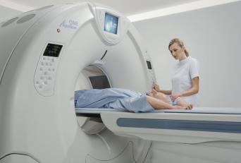
CT Scan
A CT scan (also called CAT scan), stands for Computerized Tomography. CT scans use X-rays to make detailed pictures of the internal structures of your body in cross section like slices of the inside of your body. During the test, you will lie on the table that is attached to the scanner, which is a large doughnut-shaped circular machine. The CT scanner sends X-rays through the body area being studied.
A CT scan takes between 10 – 15 minutes depending on the type of exam you will be having.
The IV contrast enhances all of the vascular structures on the images (i.e. liver, pancreas, kidneys). It will also characterize potential pathology.
Prior to the start of the CT exam the technologist will explain the procedure to you and take a brief history. After your history is reviewed, you will be positioned on the examination table. It is important for you to be comfortable, because even the slightest movement can blur the picture and result in the need for repeated scans. You will then be moved into the CT scanner. The technologist will have you in full view at all times and be in constant communication with you.
Certain exams require a special dye, called contrast, to be delivered into your body before the test starts. Contrast helps highlight certain areas on x-rays. Let your doctor know if you have ever had a reaction to contrast. You may need to take medicines before the test in order to avoid another reaction.
If contrast is used, you may also be asked not to eat or drink anything for 6 hours before the test.
If you are 60 years of age or older AND/OR you are diabetic and scheduled to receive contrast, you will be required to have a blood test to check your kidney function prior to the procedure.
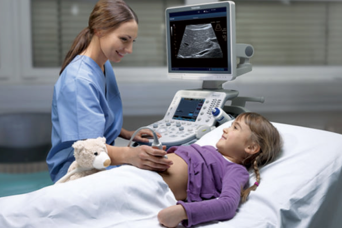
Ultrasound
The technologist (sonographer) will explain the ultrasound procedure, take a brief history, and assist you onto the examination table. A warm gel will be applied to the area of the body that will be examined. A transducer will be moved slowly over the body part being imaged. The transducer (probe) sends a signal to an on-board computer which processes the data and produces the ultrasound image. It is from this image that the diagnosis is made for the board certified radiologist to interpret and report.
Scans typically take between 20 – 30 minutes. When preparing for your exam, you should wear loose, comfortable clothing. Depending on your exam, you may have to avoid food or drink for 6 hours prior to your exam. For these exams, we try to schedule early morning appointments. There are other exams that will require you to have a full bladder prior to your exam. BASC Imaging will review all preparation instructions at the time of scheduling.
Ultrasounds are safe and noninvasive. Ultrasounds do not produce radiation.
Request an Imaging Appointment
Do you need an MRI, CT Scan or Ultrasound? Have a standing order from a physicians?
Click on the button below to schedule an appointment at BASC Imaging.
Are you a Physician?
Referring patients to BASC Imaging is easy.
Simply call our scheduling line to speak with one of our staff members and we will get your patient in as quickly as possible for an appointment. We offer referring providers the ability to see images for their patients once they have been scanned. A report will be faxed within 24 – 48 hours to the ordering provider’s office.
To register for access to the Ambra Health portal, please contact BASC Imaging to set up your account and download the Ambra Health app in the App Store.
BASC Imaging is proud to work with Board Certified radiologists from New England Baptist and Southern New Hampshire Radiology for reading services. Referring physicians can expect to receive reads within 24-48 hours of the patient’s appointment. Stat reads are available upon request.